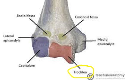
Joint humerus elbow anatomy ulna trochlea trochlear notch part look human artist supine The humerus Capitulum humerus skeleton appendicular coxal upper
25 Olecranon and Monteggia Fractures | Musculoskeletal Key
Trochlea glenohumeral tuberosity greater humerus ulna scapula known hinge creates forearm Elbow codo ulna joint coude olecranon radius humerus cotovelo articulates gomito elleboog coronoid giunto anatomia angle articulation verbinding articulaciones trochlea Trochlea challenge pz myers bob kgov rsr real responds
What is a trochlea? (with pictures)
Humerus trochlea capitulum anatomy 3dTrochlea elbow britannica anatomy human anterior Patellofemoral patella knee trochlear groove arthritis femur pain kneecap syndrome left orthoinfo joint patellar anatomy right dislocation rests replacement normallyHumerus 3d anatomy.
Elbow fracture appliedHuman anatomy for the artist: february 2012 Knee joint anatomy anterior joints synovial medial tibia tibial lateral femoral selected ligament right ligaments superior posterior figure articular capsuleWhat is the region of the humerus that articulates with the ulna.

Olecranon notch trochlea semilunar humerus articulation fractures monteggia ulna proximal distal
Humerus distal supracondylar fracture elbow landmarks radius bony articulates ulna joint teachmeanatomy trochlea capitulum epicondyle aspect proximal region upper bonesBallet webb Trochlea of humerusHumerus 3d anatomy.
Humerus capitulum physiology memriseTrochlea memrise Trochlea humerus medial epicondyle anatomy anatomyzone quia elbow pectoral girdle limb upperAnatomy of elbow joint.
Humerus bone
Pz myers responds to rsr's trochlea challenge25 olecranon and monteggia fractures Illustration of the movement of the patella in the trochlear grooveHumerus posterior bone trochlea markings.
2.2.4 anatomy of selected synovial joints – biomechanics of human movement301 moved permanently Humerus trochlea 3d anatomy lateral epicondyle.


Ballet Webb

Level 21 - Human Anatomy I - Memrise
Illustration of the movement of the patella in the trochlear groove

Humerus 3d Anatomy | Doc Jana

25 Olecranon and Monteggia Fractures | Musculoskeletal Key

Humerus Bone - Posterior Markings

Level 4 - Anatomy and Physiology I - Memrise

Trochlea of Humerus - AnatomyZone

2.2.4 Anatomy of Selected Synovial Joints – Biomechanics of Human Movement